Understanding PET-CT Scans in Cancer Diagnosis
A PET-CT scan gives your oncologist a detailed map of where cancer may be active in your body and whether it has spread. This guide explains what the scan shows, how to prepare, and what your results mean for your treatment plan.
Updated on Jul 25, 2026
Understanding PET-CT Scans in Cancer Diagnosis
A PET-CT scan combines two types of imaging in a single appointment to help your oncologist see where cancer may be growing, how active those cells are, and whether the disease has spread. If your doctor has ordered one, or if you are trying to understand results you have already received, this guide explains what happens and why it matters.
Quick answer: A PET-CT scan works by injecting a small amount of radioactive sugar into your bloodstream. Cancer cells absorb more sugar than healthy cells, so they appear as bright spots on the image. Your oncologist uses this to determine your cancer stage and plan treatment.
What Is a PET-CT Scan?
PET stands for positron emission tomography. CT stands for computed tomography. A combined PET-CT machine runs both scans in a single session, usually taking 20 to 40 minutes of active scanning.
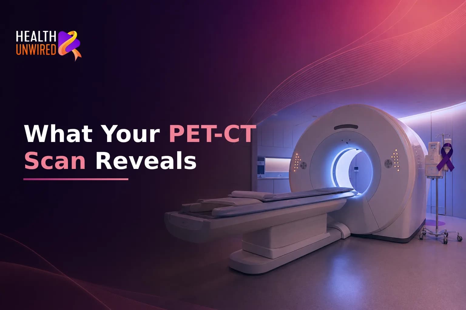
The CT part creates a detailed image of your body's structure - your bones, organs, and soft tissue. Think of it as a precise, three-dimensional X-ray. The PET part does something different: it measures how much sugar (glucose) cells in your body are absorbing.
Before your scan, a technician injects a small amount of radioactive glucose into a vein in your arm. This substance is called a tracer. The most common tracer used in cancer imaging is FDG, short for fluorodeoxyglucose. According to the American Cancer Society, a PET scan shows whether cells in a specific area are metabolically active in a way that may indicate cancer.
When the CT and PET images are layered together on a computer screen, your oncologist and radiologist can see both the exact location of a suspicious area and how the cells there are behaving. This combination gives more information than either scan alone.
What Does Your Oncologist Actually See?
Reading a PET-CT scan requires specialist training. A nuclear medicine radiologist reviews the images and writes a detailed report. Your oncologist then uses that report to decide on care. Here is what they are both looking for.
Hot spots. Areas where cells absorbed more tracer than expected appear as bright zones on the PET image. Doctors call these hot spots. They can point to cancer, but they can also show up with infections, inflammation, and some benign conditions. A trained radiologist considers the shape, location, and intensity of each hot spot alongside your medical history before drawing conclusions.
Standardized uptake value (SUV). This is a number the software calculates for each area of interest. A higher SUV generally means that cells in that area are more metabolically active. Oncologists use SUV as one piece of information when assessing how active a tumor appears and whether activity has changed between scans. Research on how oncologists read FDG PET/CT shows how the metabolic findings from the PET combine with the structural detail from the CT to create a complete clinical picture.
Spread to lymph nodes or other organs. One of the most important things a PET-CT scan can show is whether cancer has moved beyond its original location. If cells carrying the tracer show high activity in lymph nodes, the liver, or distant bones, that changes the cancer stage - and the stage directly affects which treatments are considered appropriate. Studies show that PET-CT imaging may be more accurate than CT imaging alone for staging, which can directly affect treatment choices. (source: PET/CT in Oncology - Current Status and Perspectives, PMC)
Treatment response over time. PET-CT is not ordered only at diagnosis. Your care team may request a repeat scan during or after treatment to see whether therapy is working. A tumor showing reduced tracer uptake after treatment suggests the cancer cells may be less active. For patients with lung cancer, lymphoma, and certain digestive cancers, doctors often order repeat PET-CT scans as a standard part of treatment monitoring.
How Does a PET-CT Scan Affect Your Treatment Plan?
The findings from your PET-CT scan shape your cancer stage. Stage is one of the most important factors in deciding what treatment your oncologist recommends. If you want a clear explanation of how staging works, our guide to cancer staging and TNM classification covers what each stage means and how it guides treatment choices.
If the scan shows spread that was not visible on earlier images, your oncologist may change your treatment plan significantly. Surgery may no longer be an option, or radiation areas may need to be widened, or a systemic therapy may be added. These are important changes. That is why it is worth understanding what the scan showed - and asking questions if the explanation is not clear.
If you are still in the days after your diagnosis and feel overwhelmed by tests and decisions, our article on what to do in the first 72 hours after a cancer diagnosis explains what each result typically leads to next and how to approach early decisions.
How Should You Prepare for Your PET-CT Scan?
Preparation is more important for a PET-CT scan than for most other imaging tests. Because the test measures how your cells absorb sugar, your blood glucose level on the day can affect image quality. Follow these steps before your appointment.
Fast for at least 4 to 6 hours before the scan. Do not eat or drink anything except plain water during that time. The American Cancer Society notes that fasting keeps blood sugar levels stable so the tracer can be absorbed in a way that produces accurate images. (source: How to Prep for a PET Scan, American Cancer Society) Coffee, tea, juice, and gum all count as food for this purpose - even without added sugar.
Tell your care team if you have diabetes. Diabetes changes how your body manages blood sugar, which affects how the tracer behaves. Your team may adjust your fasting instructions or check your glucose level right before the injection. If your blood glucose is too high on the day, you may need to reschedule.
Drink plenty of plain water. Staying well hydrated helps your kidneys clear the tracer from your body after the scan. Unless your doctor has told you to limit your fluids for another reason, aim for extra water in the 24 hours before your appointment.
Avoid strenuous exercise for 24 to 48 hours beforehand. Physical activity causes muscles to absorb more glucose. This can create bright areas on the scan that have nothing to do with cancer and make the images harder for the radiologist to read.
Tell your team about all medications and supplements. Some drugs and herbal preparations change how your body processes glucose. Your imaging team needs this information to prepare you correctly and to read what they see accurately.
Leave metal jewelry at home. Rings, earrings, watches, and piercings can interfere with both the CT and PET images. Leave them at home rather than removing them at arrival.
The days before a PET-CT scan, and especially the wait for results, can be stressful. If you need support, talk to your care team about options available to you.
What Happens on the Day of the Scan?
When you arrive, a nuclear medicine technician will check your blood glucose level. If it is within the acceptable range, they will place a small IV line into a vein in your arm and inject the FDG tracer.
After the injection, you will wait in a quiet room for about 45 to 60 minutes. This is the uptake period - the time the tracer needs to travel through your body and be absorbed by cells. During this time, stay as still and calm as possible. Moving, talking, and even chewing can cause muscle tissue to absorb more tracer and affect image quality.
The scan itself usually takes 20 to 40 minutes. You lie still on a flat table that moves slowly through a large, ring-shaped machine. The machine does not touch you and the process is painless. Memorial Sloan Kettering Cancer Center's PET-CT patient guide notes that you can breathe normally throughout the scan and that the technician can communicate with you through an intercom if you need anything.
After the scan, the radioactive tracer leaves your body naturally, mostly through your urine. Drinking extra fluids in the hours afterward helps speed this process. Your imaging team will give you specific guidance about any precautions, especially around young children or people who are pregnant.
When Will You Get Results - and What Do They Mean?
Results are usually ready within 24 to 48 hours. A nuclear medicine radiologist reviews the scan and sends a written report to your oncologist. Your oncologist then discusses the findings with you at a scheduled appointment or by phone or video.
The report will describe any areas of abnormal tracer uptake, the size and location of suspicious spots, how findings compare to any earlier images, and the SUV values for key areas. Do not try to interpret the raw numbers on your own. A high SUV in one area means something different depending on the cancer type, the tracer used, and your full medical picture. Ask your oncologist to explain each finding - and the conclusion at the end of the report.
If the results lead to a significant change in treatment - for example, moving away from surgery toward systemic therapy - make sure you understand how the scan drove that recommendation. If you are not satisfied with the explanation, or if the plan is a major departure from what you expected, getting a second opinion is a normal and sensible step. Our article on what changes after a second opinion in cancer care explains what tends to shift when a second specialist reviews the same clinical picture.
Should You Get a Second Opinion on Your PET-CT Results?
PET-CT interpretation is a specialist skill, and findings can be read differently depending on the radiologist's experience with a particular cancer type. A hot spot assessed as benign by one team may be flagged differently by a nuclear medicine specialist at a high-volume cancer center. Because the stage from your scan directly shapes what treatments you are offered, the accuracy of that reading matters.
If you are about to make a major treatment decision based on PET-CT findings, it is reasonable to ask a second oncologist to review the scan and your full medical picture. Online consultations make this practical: you can upload your scan files and pathology reports to be reviewed by a verified oncologist, and receive a written clinical opinion - often within 48 hours and without leaving home.
When to Talk to Your Doctor
Contact your oncology or imaging team before your scan if you have any questions about preparation - especially if you take medications that affect blood sugar or if you have diabetes. After the scan, ask your oncologist to go through the full report with you, not just the conclusion. If PET-CT findings are driving a major treatment decision, make sure you understand what was seen, what was not, and whether a second specialist has reviewed the images. A second opinion on imaging is a reasonable step before committing to any significant procedure or treatment change.
This article is for general information and is not a substitute for medical advice. Always consult your oncologist or care team about your specific situation.
Similar Topics
Understanding Your Lymphoma Diagnosis and Staging
A lymphoma diagnosis involves three steps: a biopsy, a pathology report naming your exact subtype, and a stag…
HealthUnwired Team
Sep 16, 2026
Your First Week After a Prostate Cancer Diagnosis
A prostate cancer diagnosis raises immediate questions about tests, timelines, and next steps. This guide wal…
HealthUnwired Team
Sep 9, 2026
Stomach Cancer Diagnosis and Staging Explained
A stomach cancer diagnosis raises immediate questions about stage, tests, and what comes next. This guide exp…
HealthUnwired Team
Aug 22, 2026